ABSTRACT:
In this article, we will discuss about the fascinating physiology and anatomy of human heart. The human heart is a remarkable organ responsible for pumping oxygenated blood to all parts of the body. With its intricate structure and complex physiology, the heart plays a vital role in maintaining the overall health and functionality of the human body. We will discuss about the mechanism of blood circulation. We will also provide related references to understand the concept deeply.
INTRODUCTION:
The human heart serves as the central pump of the cardiovascular system, tirelessly delivering oxygen and nutrients to the body’s tissues. This remarkable organ, roughly the size of a fist, beats approximately 100,000 times a day and can pump around 2,000 gallons of blood, illustrating its vital role in sustaining life. Understanding the physiology and anatomy of the heart is essential for comprehending its profound functional capacity.
PHYSIOLOGY AND ANATOMY OF HUMAN HEART:
1. ANATOMY OF HUMAN HEART:
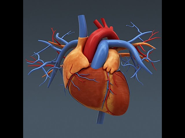
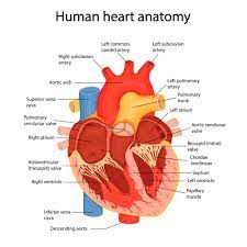
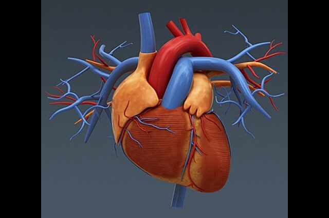
The human heart is located in the thoracic cavity, slightly left of the sternum. It enclosed within a double-layered protective sac called the pericardium. The heart comprises four chambers – two atria and two ventricles – which work together to circulate blood throughout the body. The right atrium receives deoxygenated blood through the superior and inferior vena cava. While the left atrium receives oxygenated blood from the pulmonary veins. The right ventricle pumps blood into the lungs via the pulmonary artery. While the left ventricle forcefully propels oxygenated blood into the aorta, initiating systemic circulation.
a) THE HEART WALL:
The heart composed of three distinct layers: the outer epicardium, the middle myocardium, and the inner endocardium. Richly supplied with blood vessels, the epicardium acts as a protective layer and aids in reducing friction during heart contractions. The myocardium, comprising cardiac muscle tissue, facilitates the contraction and relaxation required for pumping blood. Its extensive blood supply ensures the delivery of oxygen and nutrients essential for the cardiac muscle’s continuous energy requirements. Finally, the endocardium lines the inner surface of the heart chambers, providing a smooth surface to minimize clot formation.
b) HEART VALVES:
The heart equipped with four valves that ensure the unidirectional blood flow. The atrioventricular (AV) valves, namely the tricuspid valve on the right side and the mitral valve on the left side, separate the atria from the ventricles. These valves prevent blood backflow into the atria during ventricular contractions. Semilunar valves, including the pulmonary valve and aortic valve, situated between the ventricles and their respective arteries, preventing blood regurgitation back into the ventricles.

2. PHYSIOLOGY OF HUMAN HEART:
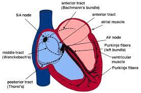
The human heart’s rhythmic contractions occur due to its specialized conduction system. The sinoatrial (SA) node, located in the right atrium, serves as the natural pacemaker, generating electrical impulses that initiate each heartbeat. These electrical signals then travel through the atria, causing them to contract and squeezing blood into the ventricles. The impulses are passed to the atrioventricular (AV) node, situated at the base of the right atrium, before being rapidly transmitted through the atrioventricular bundle (Bundle of His) and its subdivisions, known as the Purkinje fibers. This orchestrated conduction system ensures synchronized contractions of the ventricles, effectively pumping blood out of the heart.

CONCLUSION:
The human heart’s physiology and anatomy exhibit features that facilitate its vital function, tirelessly pumping blood throughout the body. Understanding the structural arrangement of the heart, its chambers, valves, and conduction system, is essential for comprehending the complexity and efficiency of this remarkable organ. By exploring the physiology and anatomy of the heart, researchers can continually advance our knowledge of cardiovascular health, leading to improved treatments and preventive measures for cardiac diseases.
REFERENCES:
Tortora, G. J., & Derrickson, B. H. (2017). Principles of Anatomy and Physiology. Wiley. https://www.wiley.com/en-ie/Principles+of+Anatomy+and+Physiology,+15th+Edition-p-9781119320647
Silverthorn, D. U. (2018). Human Physiology: An Integrated Approach. Pearson. https://www.pearson.com/nl/en_NL/higher-education/subject-catalogue/biology/Human-physiology-silverthorn-8e.html
Guyton, A. C., & Hall, J. E. (2015). Textbook of Medical Physiology. Elsevier. https://repository.poltekkes-kaltim.ac.id/1147/1/Guyton%20and%20Hall%20Textbook%20of%20Medical%20Physiology%20(%20PDFDrive%20).pdf


